Single Cell Studies and Cytoskeleton Mechanics in Quasi-3D
Osteocytes are three-dimensional, ellipsoidal, mature bone cells encased in mineralized extracellular matrix. It has been hypothesized that the dominant loading mechanism of the mechanosensing osteocytes is fluid shear stress resulting from the deformation of bone. Recent experimental and theoretical evidence suggest that the ellipsoidal cell body may be less sensitive to fluid shear than the slender cellular processes. However, for adherent cells under fluid flow, the interactions between the flow field, the cell, and the substrate are dynamic and complex. The conventional top-to-bottom view microscopy of cell deformation is insufficient to characterize the intracellular deformation of the adhered cells at their apical surface (cell body) and at their basal surface (cellular processes and dendrites) under flow.
Osteocytes are three-dimensional, ellipsoidal, mature bone cells encased in mineralized extracellular matrix. It has been hypothesized that the dominant loading mechanism of the mechanosensing osteocytes is fluid shear stress resulting from the deformation of bone. Recent experimental and theoretical evidence suggest that the ellipsoidal cell body may be less sensitive to fluid shear than the slender cellular processes. However, for adherent cells under fluid flow, the interactions between the flow field, the cell, and the substrate are dynamic and complex. The conventional top-to-bottom view microscopy of cell deformation is insufficient to characterize the intracellular deformation of the adhered cells at their apical surface (cell body) and at their basal surface (cellular processes and dendrites) under flow.
Obtaining reasonable 3D cell information using current spinning-disk confocal microscopy technologies takes approximately .5 to .8 seconds. The limitation of the z-stack scanning speed makes the confocal microscope a non-ideal choice to image events requiring a higher temporal resolution, such as the imaging of osteocytes sheared at a 1Hz oscillatory loading frequency, which require at least 8-12 frames per second to adequately cover the 1 Hz loading profile.
Our laboratory developed a novel two-microscope system that consists of an upright microscope and an inverted microscope. A 45 degree mirror is placed in the light path of the upright microscope to obtain "sideview" images. Cells placed in a square glass tube provide the necessary optics for high resolution imaging and fluid flow. Essentially, the system images two orthogonal views of a single cell at a frame rate only limited by the camera frame rate. This "quasi-3D" system has avoided the use of z-stack raster-scanning of confocal microscopy and the limitations of traditional inverted microscopy to obtain far more simultaneous cellular information than previously possible. Additional beamsplitters allow simultaneous imaging of several dyes in the same acquisition.
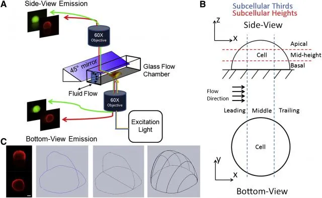
(A) Schematic of the quasi-3D microscopy system.
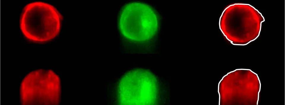
(B) Axes for bottom-view and side-view images. Subcellular location divisions are depicted. Subcellular thirds (leading, middle, and trailing) for bottom- and side-views are indicated by blue lines, and subcellular heights (apical, mid-height, and basal) for side-view are shown by red lines.
(C) Schematic of conversion of cell plasma membrane boundaries from side and bottom views to obtain a whole-cell volume. Boundary points tracked by digital image correlation are fit to a spline function and then lofted into a 3D shape. Scale bar is 5 μm.
Taken all together, this exciting technology enables simultaneous visualization and quantification, in real-time and in 3D, of the dynamics of two cytoskeletal components and signal activation via fluorescence resonance energy transfer (FRET). This unique approach provides high spatial and temporal specificity in studying single cell mechanics and mechanotransduction under complex flow conditions. Not only is this a breakthrough technology in bone cell mechanics and mechanotransduction, but also a valuable approach in cancer cell migration/motility and endothelial cell mechanobiology.
Related papers
- Baik, AD et al "Quasi-3D Cytoskeletal Dynamics of Osteocytes under Fluid Flow" Biophysical Journal 2010
- Baik, AD et al "Simultaneous tracking of 3D actin and microtubule strains in individual MLO-Y4 osteocytes under oscillatory flow" BBRC 2013
Using this technology, we have demonstrated contractions of the actin cytoskeleton that correspond to rises in intercellular Ca2+ levels. Continuing work aims to further elucidate the mechanism of contraction and the coordination of Ca2+ signaling and actin dynamics in response to flow.