Research Areas
The Bone Bioengineering Laboratory performs multiscale research in two main areas: clinical mechanics and mechanobiology. The BBL has also developed cutting edge software called Individual Trabeculae Segmentation (ITS) , which is used in both our work and others for rapid and novel analysis of trabecular bone parameters.
The clinical mechanics group studies the affect of both morphology and mechanical properties of individual trabeculae on whole bone properties. This is accomplished through use of cutting-edge imaging technologies and advanced software to study microstructural changes in bone as well as customized mechanical loading devices that allow mechanical properties to be derived at multiple scales.
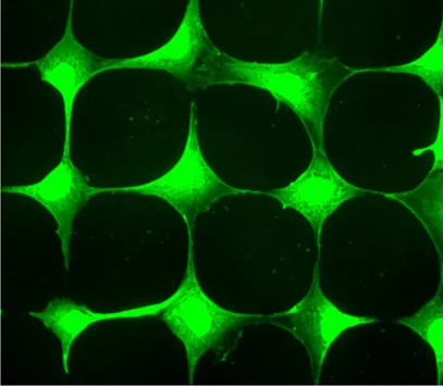
The mechanobiology group studies the mechanisms of osteocyte mechanosensation and mechanotransduction. We have developed novel in vitro, ex vivo, and in vivo technologies that allow the study of osteocyte mechanobiology at several scales - from the immediate response of single cells to mechanical stimulus all the way through effects on long-term, whole bone response to loading when osteocyte function is impaired.
Visit each area's respective page to read a high-level description and learn about specific projects in that area.
Clinical Mechanics
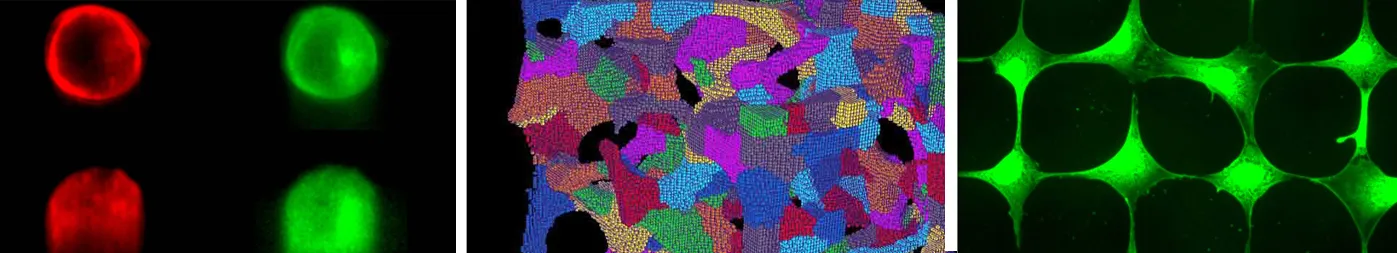
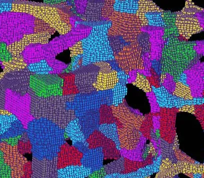
The clinical mechanics group studies the affect of both the morphology and mechanical properties of individual trabeculae on whole bone properties. This is accomplished through the use of cutting-edge imaging technologies and advanced software to study microstructural changes in bone as well as customized mechanical loading devices that allow mechanical properties to be derived at multiple scales. A centerpiece of our clinical mechanics imaging projects is the use of a novel, BBL-developed software called Individual Trabeculae Segmentation (ITS) , which enables images of trabecular bone to be accurately and rapidly decomposed into plates and rods and unique trabecuale parameters, such as thickness, orientation, and number, to be directly measured. Using all of these techniques, the clinical mechanics group can detect subtle changes in bone microarchitecture as potential markers of pathological bone conditions and relate these changes to changes in bone mechanical properties that can lead to, for example, increased fracture risk.
Mechanobiology
The osteocyte is a long-lived, highly dendritic cell forming a vast, interconnected network within the mineralized bone matrix. It has only been over the last couple of decades that cell isolation and imaging techniques have advanced enough to allow deeper study of this most interesting cell. The osteocyte is acknowledged as integral to sensation of changing mechanical forces at the whole bone level (mechanosensation) and has also been identified as the major source of key proteins that modulate osteoblast and osteoclast response to loading (cells that build and resord bone, respectively). However, the mechanisms by which mechanosensation translates to changes in modulating proteins (mechanotransduciton) as well as how these proteins are transported from the osteocyte, embedded within the bone matrix, to the effector cells at the surface and in the marrow space of the bone is poorly understood.
ITS and FEA of skeletons
In the area of image based microstructural and finite element analyses of human skeletons, we have innovated and advanced an individual trabeculae segmentation (ITS) technique and demonstrated its unique power in basic micromechanics of human trabecular bone. More importantly, we have successfully translated this ITS technology into clinical studies of metabolic bone diseases. The most exciting development is the recent breakthrough in identifying dramatic and striking microstructural differences in bones between Chinese-Americans and Caucasians. It will have significant clinical, basic science, and anthropological implications as we begin to explore the genetic and environmental causes of these remarkable differences. Projects in this area are listed below.